Anatomical Models
-
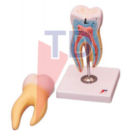 |
HUMAN TOOTH MOLAR 15 TIMES |
| Code:PA-60 |
Molar tooth dissectable into 2 parts, showing the internal structure, longitudinal section through crown 2 roots and pulp cavity. Mounted on stand. Numbered with English key card.
|
-
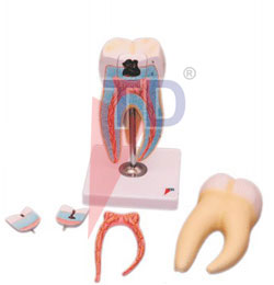 |
UPPER TRIPLE ROOT MOLAR WITH CARIES 15 TIMES FULL SIZE 6 PARTS |
| Code:PA-63 |
Longitudinal section through crown, 2 roots and pulp cavity. Removable pulp and three tooth inserts with different stages of advanced caries. On stand. Numbered with English key card.
|
-
| HUMAN TOOTH |
| Code:PA-64 |
A set of 5 models dissectable into 2 parts each, showing the detailed structure of incisor, canine, premolar, lower & upper molar tooth on stand.
|
 |
-
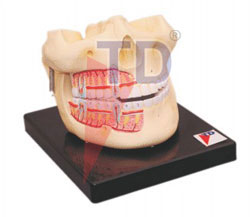 |
HUMAN UPPER AND LOWER JAW |
| Code:PA-69 |
A model showing various positions of teeth in the upper and lower jaws. Internal wall is removed to show the incisors, canine premolars, molars in full view to show the tooth roots, spongise, vessels and nerves serving them. Dissectable into 2 parts. Mounted on base. Numbered with English key card.
|
-
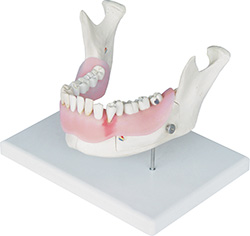 |
DENTAL DISEASE ANATOMY |
| Code:PA-72 A |
This model shows illustration of a lower jaw with 16 removable teeth of an adult magnified two times. One half of the model shows eight healthy teeth and healthy gums, and the other half of the model shows following dental diseases:
• Dental Plaque
• Dental calculus (tartar)
• Inflammation of the root
• Periodontitis
• Fissure, approximal and smooth surface caries
One part of the front bone section can be removed to view the roots, vessels and nerves. Two molars are sectioned along the length to show the inside of the tooth..
|
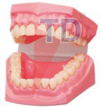 |
DENTAL CARE MODEL |
| Code:PA-75 |
This model is a demonstration model used by the students to study the teeth.
|
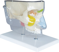 |
NOSE WITH PARANASAL SINUSES, 5-PARTS |
| Code:PA-77 A |
This model illustrates the structure of the nose with the paranasal sinuses in the upper right half of a face in 1.5 times enlargement. The following structures can be seen from outside, differentiated by color:
• The outer nasal cartilages
• The nasal, maxillary, frontal and sphenodial sinuses
• The opened maxillary sinus when the zygomatic arch is removed
The following structures are shown in a median section:
• The nasal cavity, lined with mucosa, with the nasal conchae
• The arteries of the mucous membrane
• The olfactory nerves
• The innervation of the lateral wall of the nasal cavity, the nasal conchae and the roof of mouth (palate)
|

